What is the optic chiasm function
 Rachel Young
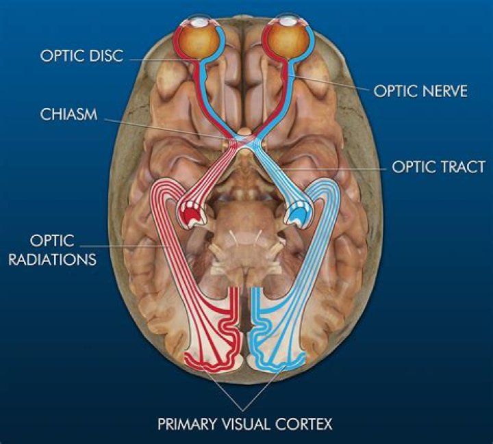
 The optic chiasm or optic chiasma is an X-shaped space, located in the forebrain, directly in front of the hypothalamus. Crucial to vision, the left and right optic nerves intersect at the chiasm, thus creating the hallmark X-shape.
What is optic chiasm quizlet?
Optic chiasm. Where the optic nerves meet in the 3rd ventricle of the brain. Here is where visual information from the right and left eye cross to the contralateral side of the visual systems. It is also ensheathed with the meninges and is surrounded by CSF.
Where is the optic chiasm located quizlet?
is the part of the brain where the optic nerves (CN II) partially cross. The optic chiasm is located at the bottom of the brain immediately below the hypothalamus.
What happens if optic chiasm is damaged?
Damage to the retina or one of the optic nerves before it reaches the chiasm results in a loss of vision that is limited to the eye of origin. In contrast, damage in the region of the optic chiasm—or more centrally—results in specific types of deficits that involve the visual fields of both eyes (Figure 12.8).What is the optic tract?
The optic tract is a bundle of nerve fibers that serves to carry visual information from the optic chiasm to the left and right lateral geniculate bodies as a part of the visual pathway.
What causes optic chiasm lesions?
Lesions that may compress the visual chiasm include pituitary adenoma, craniopharyngioma, and meningioma. Of these, pituitary adenoma is the third most common intracranial tumour in Japanese national statistics, and it is also common in other countries, accounting for 12%–15% of all intracranial tumors6–8.
How does the optic chiasm contribute to human vision?
The partial crossing over of optic nerve fibres at the optic chiasm allows the visual cortex to receive the same hemispheric visual field from both eyes. Superimposing and processing these monocular visual signals allow the visual cortex to generate binocular and stereoscopic vision.
Where is the optic chiasm located in relation to the pituitary gland quizlet?
The optic chiasm is located at the bottom of the brain immediately below the hypothalamus. on the ventral side of brain under optic chiasm and pituitary location.What happens to the optic nerve at the optic chiasm?
At a structure in the brain called the optic chiasm, each optic nerve splits, and half of its fibers cross over to the other side. Because of this anatomic arrangement, damage along the optic nerve pathway causes specific patterns of vision loss.
What part of the retina lacks photoreceptors?blind spot, small portion of the visual field of each eye that corresponds to the position of the optic disk (also known as the optic nerve head) within the retina. There are no photoreceptors (i.e., rods or cones) in the optic disk, and, therefore, there is no image detection in this area.
Article first time published onWhich of the following statements best describes trichromatic theory and opponent process theory group of answer choices?
Which of the following statements best describes trichromatic theory and opponent-process theory? Research has not supported either theory. Both theories are equally accurate, but they apply to different levels of the nervous system. The trichromatic theory is more accurate than the opponent-process theory.
Where do optic tracts go to?
The optic tract is an important part of the visual pathway. Almost all of the axons of the left and right and left optic tracts synapse with the cells of the ipsilateral lateral geniculate nucleus. The efferent fibres emerge as the optic radiation and ultimately travel to the primary visual cortex.
Is the optic tract before the optic chiasm?
The optic tract (from the Latin tractus opticus) is a part of the visual system in the brain. It is a continuation of the optic nerve that relays information from the optic chiasm to the ipsilateral lateral geniculate nucleus (LGN), pretectal nuclei, and superior colliculus.
Where do optic tracts exist?
The optic tract is an extension of the optic nerve located in the brain. It begins at the area where information from the left eye and right eye cross (or “decussate”) to create a complete visual picture.
What is a chiasm disorder?
Chiasmal syndrome is the set of signs and symptoms that are associated with lesions of the optic chiasm, manifesting as various impairments of the sufferer’s visual field according to the location of the lesion along the optic nerve.
What causes bitemporal hemianopia?
A bitemporal hemianopia is almost always caused by damage to the optic chiasm and can occur from the direct or indirect effects of a variety of lesions, including tumors,1 aneurysms,2 and, less frequently, inflammatory and ischemic diseases.
How does bitemporal hemianopia occur?
Bitemporal hemianopsia most commonly occurs as a result of tumors located at the mid-optic chiasm. Since the adjacent structure is the pituitary gland, some common tumors causing compression are Pituitary adenomas, and Craniopharyngiomas. Also another relatively common neoplastic etiology is Meningiomas.
What does oculomotor nerve do?
The oculomotor nerve is the third cranial nerve (CN III). It allows movement of the eye muscles, constriction of the pupil, focusing the eyes and the position of the upper eyelid. Cranial nerve III works with other cranial nerves to control eye movements and support sensory functioning.
Is the fovea responsible for central vision?
The fovea is responsible for sharp central vision (also called foveal vision), which is necessary in humans for activities for which visual detail is of primary importance, such as reading and driving.
Where is the optic chiasm located in relation to the hypothalamus?
The optic chiasm or optic chiasma is an X-shaped space, located in the forebrain, directly in front of the hypothalamus. Crucial to vision, the left and right optic nerves intersect at the chiasm, thus creating the hallmark X-shape.
What produces oxytocin and ADH?
The paraventricular nuclei produce the hormone oxytocin, whereas the supraoptic nuclei produce ADH. These hormones travel along the axons into storage sites in the axon terminals of the posterior pituitary.
What holds the pituitary gland?
Within your skull, there’s a small, bony nook at the base of your brain that holds and protects your pituitary gland (which controls how hormones work in your body). This tiny structure is called the sella turcica.
What happens when light strikes a photoreceptor?
When light hits a photoreceptor, it causes a shape change in the retinal, altering its structure from a bent (cis) form of the molecule to its linear (trans) isomer.
Which eye feature provides vitamin A for photoreceptors?
Rods are a type of photoreceptor cell present in the retina that transmits low-light vision and is most responsible for the neural transmission of nighttime sight. Rods have a singular photopigment, rhodopsin, which utilizes the protein scotopsin and the Vitamin A-derived cofactor, retinol.
Which part of the retina is responsible for the sharpest vision?
In the center of the retina is the macula, which is responsible for our near vision. In the center of the macula is the fovea, responsible for our sharpest vision.
What term describes the continuation of a visual sensation after removal of the stimulus?
An afterimage describes the continuation of a visual sensation after removal of the stimulus. For example, when you stare briefly at the sun and then look away from it, you may still perceive a spot of light although the stimulus (the sun) has been removed.
How does a cochlear implant enable the deaf to hear quizlet?
How does a cochlear implant enable the deaf to hear? It receives incoming sound information and directly stimulates the auditory nerve to transmit information to the brain.
What has research by goolkasian & Woodbury 2010 demonstrated about pattern perception?
What has research by Goolkasian & Woodbury (2010) demonstrated about pattern perception? Subliminal priming is more effective than priming above the absolute threshold. … Those who receive less auditory priming are more likely to hear things than those who receive more auditory priming.
What happens if you cut the left optic tract?
Damage at site #1: this would be like losing sight in the left eye. The entire left optic nerve would be cut and there would be a total loss of vision from the left eye.
Why is optic nerve a tract?
The trabeculae form a crisscross pattern outlining “pores” through which the nerve fiber bundles pass. The myelinated orbital portion of the optic nerve can be considered more a tract of the brain than a true cranial nerve.
What supplies the optic tract?
The blood supply of the optic tract is supplied by thalamic perforators of the posterior cerebral artery and branches of the anterior choroidal artery off the internal carotid artery. Each optic tract contains fibers from the contralateral hemifield.