What diseases cause cotton wool spots
 Christopher Martinez
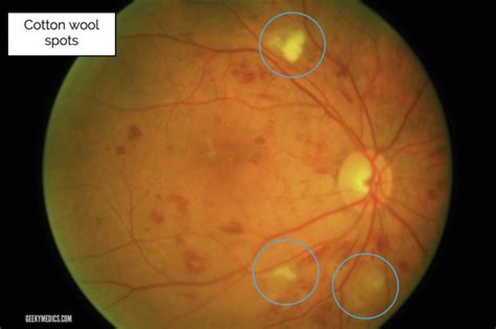
 Cotton-wool spots (CWSs) are common retinal manifestations of many diseases including diabetes mellitus, systemic hypertension, and acquired immunodeficiency syndrome. Clinically they appear as whitish, fluffy patches on the retina and eventually fade with time.
What causes cotton wool bodies?
Cotton-wool spots are composed of accumulations of neuronal debris within the nerve fibre layer. They result from ischaemic disruption of nerve axons, the swollen ends of which are known as cytoid bodies, seen on light microscopy as globular structures in the nerve fibre layer ( Fig. 13.11A ).
What are cotton-wool spots on retina?
Cotton-wool spots (CWS) (Figure) are acute signs of vascular insufficiency to an area of retina. They have been described in many conditions, but only occasionally cause symptoms in patients. The most common symptoms associated with retinal CWS can include scotoma, arcuate defects, blurred vision, and amaurosis fugax.
Can lupus cause cotton-wool spots?
Mild lupus retinopathy consists of cotton–wool spots, perivascular hard exudates, retinal hemorrhages, and vascular tortuosity [7]. Moderate lupus retinopathy has focal or generalized arteriolar constriction and venous tortuosity.How does diabetes cause cotton-wool spots?
Due to the strong association with microangiopathic diseases such as diabetes mellitus and systemic hypertension, the interruption in axoplasmic flow in cotton-wool spots is thought to result from focal ischemia associated with occlusion of precapillary arterial flow, though other possible etiologies have also been …
Are cotton wool spots normal?
In otherwise healthy patients, the observance of a cotton wool spot (CWS) is not considered normal. A single cotton wool spot in one eye can be the earliest ophthalmoscopic finding in diabetic or hypertensive retinopathy.
Are cotton wool spots serious?
Cotton-wool spots are tiny white areas on the retina, the layer of light-sensing cells lining the back of the eye. Caused by a lack of blood flow to the small retinal blood vessels, they usually disappear without treatment and do not threaten vision. They can, however, be an indication of a serious medical condition.
Can you see lupus in the eyes?
People with lupus can get retinal vasculitis, which limits the blood supply to the retina, which can have significant effects on vision. The eye then attempts to repair itself, but when the retina tries to repair itself it forms new blood vessels which can form in areas of the eye that can impair vision.What are the typical signs and symptoms of autoimmune diseases using lupus as an example?
- Fatigue.
- Fever.
- Joint pain, stiffness and swelling.
- Butterfly-shaped rash on the face that covers the cheeks and bridge of the nose or rashes elsewhere on the body.
- Skin lesions that appear or worsen with sun exposure.
Microaneurysms appear as grape-like or spindle-shaped dilations of retinal capillaries on light microscopy. They can be either hypercellular or acellular. By ophthalmoscopic examination, microaneurysms appear as tiny, intraretinal red dots located in the inner retina.
Article first time published onCan cotton wool spots be treated?
Cotton-wool spots are tiny white areas on the retina, the layer of light-sensing cells lining the back of the eye. Caused by a lack of blood flow to the small retinal blood vessels, they usually disappear without treatment and do not threaten vision.
Do cotton wool spots cause headaches?
We have observed three cases of isolated cotton-wool spot (CWS) accompanied by a history of migraine, and would suggest a pathophysiological association between these two clinical entities. All three patients presented with a new visual disturbance associated with recent migraine.
Is retinopathy a disease?
Retinopathy means disease of the retina. There are several types of retinopathy but all involve disease of the small retinal blood vessels. Signs of retinopathy (see photograph) can be seen when the retina is viewed through the pupil with an ophthalmoscope.
What is Purtscher's retinopathy?
Background. Purtscher retinopathy is a hemorrhagic and vasoocclusive vasculopathy, which, in 1912, was first described as a syndrome of sudden blindness associated with severe head trauma. These patients had findings of multiple white retinal patches and retinal hemorrhages that were associated with severe vision loss.
What are the stages of diabetic retinopathy?
The three stages of NPDR are mild, moderate, and severe, which may progress to the other type, or fourth stage, proliferative diabetic retinopathy.
What is retinopathy diabetes?
Diabetic retinopathy (die-uh-BET-ik ret-ih-NOP-uh-thee) is a diabetes complication that affects eyes. It’s caused by damage to the blood vessels of the light-sensitive tissue at the back of the eye (retina).
What is cotton wool disease?
Cotton wool disease, also known as saddleback, fin rot and black patch necrosis, are all descriptive names for the same bacteria, columnaris (Flavobacterium columnare). This bacteria is commonly mistaken for a fungus, given its pale color and raised appearance.
What causes myelinated nerve fibers in eye?
Summary. Myelinated nerve fiber layer (mNFL) is a benign clinical entity that results from an embryologic developmental anomaly whereby focal areas of the retinal nerve fiber layer fail to lose their myelin sheath. Clinically, mNFL appears as distinct white patches on the inner retinal surface.
Do retinal hemorrhages go away?
Retinal hemorrhages, especially mild ones not associated with chronic disease, will normally reabsorb without treatment. Laser surgery is a treatment option which uses a laser beam to seal off damaged blood vessels in the retina.
Why does neovascularization occur?
Corneal neovascularization happens when new blood vessels come into the cornea from an area of the eye called the limbus. The new blood vessels can cause inflammation and scarring that affect your vision.
Do Roth spots go away?
There’s no specific treatment for Roth spots, since a variety of conditions can cause them. However, once the underlying condition is treated, Roth spots usually go away on their own.
What are the top 5 signs of lupus?
- loss of appetite, nausea, vomiting, diarrhea, and weight loss.
- shortness of breath.
- joint inflammation, stiffness, and pain.
- swollen glands.
- muscle pain.
- chest pain when you take a deep breath.
- hair loss.
- sun sensitivity.
What are the top 10 signs of lupus?
- Achy or swollen joints (arthralgia)
- Unexplained fever (more than 100° F)
- Swollen joints (arthritis)
- Prolonged or extreme fatigue.
- Skin rash, including a butterfly-shaped rash across the cheeks and nose.
- Pain in the chest when breathing deeply (pleurisy)
- Hair loss.
What are the 3 types of lupus?
There are three types: Acute cutaneous lupus. Chronic cutaneous lupus erythematosus, or discoid lupus erythematosus (DLE) Subacute cutaneous lupus erythematosus.
Does lupus cause weight gain?
Weight changes — Lupus can sometimes cause weight loss or weight gain. Weight loss may be unintentional and due to decreased appetite or problems with the digestive system (see ‘Digestive system’ below). It can also be a side effect of some medications used to treat lupus.
What does lupus fatigue feel like?
Fatigue with Lupus. Fatigue is defined as feeling tired or lacking energy, no matter how well or how long you sleep. This exhaustion can be both physical and mental. Some people describe it as a similar feeling to having the flu.
Can lupus affect your bowel?
This makes digestive issues extremely common in people with lupus though usually not serious. Lupus can attack any part of the GI system, including the stomach, intestines, liver, pancreas, bile ducts, gallbladder, and esophagus.
What is micro aneurysm?
Microaneurysms are tiny outpouchings of blood that protrude from an artery or vein. When they occur in the eye, they are known as retinal microaneurysms. If these protrusions open, they leak blood into the tissues of the retina.
How are Microaneurysms treated?
Treatments like a change in diet and exercise can help manage blood sugar levels and keep blood vessels in the eye from being damaged. Additionally, once blood vessels in the eye start leaking, laser surgery can help cauterize them and stop or slow the leaking.
Can you see aneurysm in eye exam?
A Brain Aneurysm They will check to see if you have increased eye pressure, retinal bleeding, swelling of the optic nerve, and/or drooping eyelids. Tell your eye doctor if you experience crossed eyes, as this is may be a symptom of an aneurysm or a stroke.
Do hard exudates go away?
These plaques often cause significant visual loss when deposited in the foveal region. Until now, there have been no treatment guidelines for this pathology, and unfortunately, hard exudates have often gone unresolved with scant or no recovery for the patient.