Where is the Endothoracic fascia
 Christopher Lucas
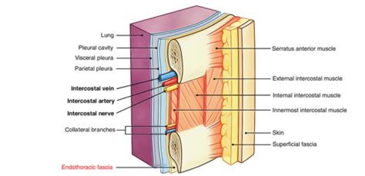
 The endothoracic fascia forms a connective tissue layer between the inner aspect of the chest wall and the costal parietal pleura.
What is Endothoracic?
The endothoracic fascia is the layer of loose connective tissue deep to the intercostal spaces and ribs, separating these structures from the underlying pleura. This fascial layer is the outermost membrane of the thoracic cavity. … It separates the internal thoracic artery from pleura.
Does thorax have deep fascia?
The chest wall has 10 layers, namely (from superficial to deep) skin (epidermis and dermis), superficial fascia, deep fascia and the invested extrinsic muscles (from the upper limbs), intrinsic muscles associated with the ribs (three layers of intercostal muscles), endothoracic fascia and parietal pleura.
What is Endoabdominal fascia?
Parietal abdominal fascia (endoabdominal fascia) may be the fascia that covers the abdominal cavity, or a generic term including extraperitoneal and visceral fascia. … Transversalis fascia is the inner epimysium of transversus abdominis muscle; no separate deep investing fascia exists.What is a deep fascia?
Deep fascia is a dense connective tissue that is commonly arranged in sheets that form a stocking around the muscles and tendons beneath the superficial fascia (1). … The superficial fascia has two layers: the external fatty layer and the deep membranous layer (2,3).
Where is thoracic cavity?
The thoracic cavity is found deep to the thoracic wall, superior to the diaphragm, and inferior to the root of the neck (thoracic aperture).
Where is the internal intercostal membrane?
The internal intercostal membrane is the continuation of the internal intercostal muscles at the vertebral end of the intercostal spaces.
Where is the posterior wall located?
Extends from the 12th rib to the iliac crest. Laterally goes to the internal oblique and transversus abdominis muscles.Where is the Transversalis fascia?
The transversalis fascia is a thin layer of connective tissue lining most of the abdominal cavity between the posterior surface of the transversus abdominis and superficial to the extraperitoneal fat and peritoneum.
Is fascia the same as peritoneum?Parietal peritoneum (or fascia): this layer is a thin serous membrane acting as a balloon which lines the abdomen and into which the organs are pressed into from the outside. Visceral peritoneum (or layer): this layer lines the organs.
Article first time published onWhere is fascia located in the body?
In short, fascia is connective tissue. It surrounds body parts from organs to muscles to blood vessels. It can also be a tough part of the body on its own, like the thick plantar fascia that stabilizes the arch on the bottom of the foot.
Where is fascia in skeletal muscle?
Fascia, connective tissue outside the epimysium, surrounds and separates the muscles.
What is a fascia in anatomy?
Introduction. Fascia is made up of sheets of connective tissue that is found below the skin. These tissues attach, stabilize, impart strength, maintain vessel patency, separate muscles, and enclose different organs.
What are the 3 types of fascia?
Fascia is classified by layer, as superficial fascia, deep fascia, and visceral or parietal fascia, or by its function and anatomical location.
Is deep fascia present in abdomen?
Deep Scarpa’s fascia, which is a thinner and denser membranous layer overlying the muscle layer of the abdominal wall. … It is firmly attached to the linea alba and pubic symphysis and fuses with the fascia lata (deep fascia of the thigh) right below the inguinal ligament.
How many fascias are in the back?
Thoracolumbar fasciaLatinfascia thoracolumbalis, fascia lumbodorsalisTA98A04.3.02.501TA22242FMA25072
Where are the external and internal intercostal muscles located?
The internal intercostal muscles (in the inside of the ribcase) extend from the front of the ribs, and go around back, past the bend in the ribs. The external intercostal muscles (on the outside of the ribcase) wrap around from the back of the rib almost to the end of the bony part of the rib in front.
What part of the body do the intercostal muscles attach to?
The intercostal muscles are a group of muscles found between the ribs which are responsible for helping form and maintain the cavity produced by the ribs. The muscles assist with expansion and contraction during breathing.
What is intercostal pain?
Intercostal neuralgia is nerve pain that affects the area below your ribs and can be caused by several different conditions. People with intercostal neuralgia experience a lot of pain in their ribs, chest, or upper abdominal area.
What organ is in the middle of your chest?
The heart and the lungs reside in the thoracic cavity, as well as many blood vessels. The inner organs are protected by the rib cage and the sternum.
What are thoracic organs?
The organs of the thorax include the thymus gland, the breasts, the heart, the lungs, the tracheobronchial tree and the pleurae. The thymus gland is located in the superior mediastinum of the thoracic cavity but may also extend into the neck.
What are the compartments of thoracic organs?
Compartments of the mediastinumStructureOrgansSuperior mediastinumThymus (prepubertal) Trachea EsophagusInferior mediastinumAnterior mediastinumRemnants of the thymus (postpubertal) Connective tissue and fatMiddle mediastinumHeart and pericardium Tracheal bifurcation and the left and right main bronchi
Where does the Transversalis fascia come from?
The transversalis fascia (or transverse fascia) is a thin aponeurotic membrane of the abdomen. It lies between the inner surface of the transverse abdominal muscle and the parietal peritoneum. It forms part of the general layer of fascia lining the abdominal parietes.
Is fascia Transversalis a deep fascia?
Transversalis fascia is the inner epimysium of transversus abdominis muscle; no separate deep investing fascia exists.
Which covering of testis is formed by fascia Transversalis?
The spermatic fascia is a bilayered fascia covering the testis; both layers are derived from abdominal muscle or fascia.
Is the psoas anterior or posterior?
The psoas major unites with the iliacus at the level of the inguinal ligament. It crosses the hip joint to insert on the lesser trochanter of the femur. The iliopsoas is classified as an “anterior hip muscle” or “inner hip muscle”.
Do females have Colles fascia?
From the scrotum it may be traced backward into continuity with the deep layer of the superficial fascia of the perineum (superficial perineal fascia or fascia of Colles). In the female, it is continued into the labia majora and from there to the fascia of Colles.
What organ is posterior to the stomach?
The posterior surface of the stomach is related to the left hemidiaphragm, the spleen, the left kidney (and adrenal), and the pancreas (stomach bed).
Where is the left anterior abdominal wall?
This is a small triangular muscle, found superficially to the rectus abdominis. It is located inferiorly, with its base on the pubis bone, and the apex of the triangle attached to the linea alba.
Where is the anterior abdominal wall?
The muscles of the anterior abdominal wall are located near the midline between the costal margin superiorly and the pubis inferiorly. There are two pairs of muscles, each located immediately lateral to the linea alba. The majority of the anterior abdominal wall is formed by the rectus abdominis muscle.
What ligaments are in your abdomen?
These abdominal ligaments are mostly named according to the structures they hold. They include the suspensory ligaments of the liver (right and left triangular ligaments, falciform ligament) and the peritoneal ligaments of the stomach (splenorenal ligament, gastrosplenic ligament, greater omentum, and lesser omentum.