What leads Makeup The einthovens triangle
 William Burgess
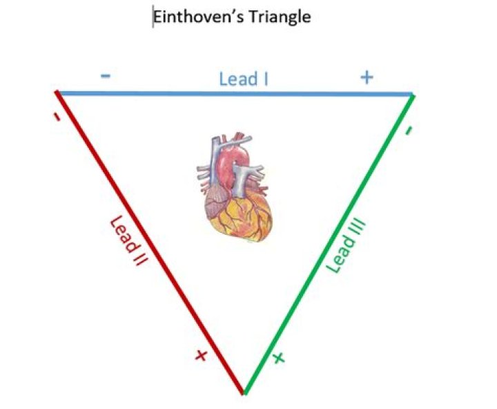
 Einthoven’s triangle is an imaginary formation of three limb leads in a triangle used in electrocardiography, formed by the two shoulders and the pubis. The shape forms an inverted equilateral triangle with the heart at the center. It is named after Willem Einthoven, who theorized its existence.
Which side of einthoven's triangle is formed by lead I?
Lead I extends horizontally from the right arm to the left arm, with the actual left arm electrode being the positive pole. Lead II forms one of the vertical arms of the triangle and stretches from the right hand to the left leg with the electrode positioned on the left leg being the positive pole.
What plane does the 3 lead ECG placement einthoven's triangle view the heart in?
Leads I, II, III, aVF, aVL and aVR are all derived using three electrodes, which are placed on the right arm, the left arm and the left leg. Given the electrode placements, in relation to the heart, these leads primarily detect electrical activity in the frontal plane.
What are the 3 bipolar leads?
The bipolar extremity leads are called I, II and III. The unipolar extremity leads are called avR, avL and avF, and the chest leads are called V1–V6.What lead is created between the right and left arm?
Lead I represents the potential difference between the right and left arm; an electrical impulse moving from right to left generates a positive ECG deflection in this lead. Lead II is the potential difference between the right arm and leg; a positive ECG deflection occurs when the impulse direction is toward the leg.
Why is lead 2 the standard lead?
To assess the cardiac rhythm accurately, a prolonged recording from one lead is used to provide a rhythm strip. Lead II, which usually gives a good view of the P wave, is most commonly used to record the rhythm strip.
What is einthoven's triangle and what does the axis refer to?
what is Einthoven’s triangle and what does the axis refer to? it’s an imaginary equilateral triangle formed by the three standard leads. it refers to the average direction that electricity moves through the heart. Lead aVR. voltage difference between right arm and midpoint of left arm and left leg.
Why is aVR inverted?
The aVR is often neglected lead. It is an unipolar lead facing the right superior surface. As all the depolarisations are going away from lead aVR, all waves are negative in aVR (P, QRS, T) in normal sinus rhythm.Why is einthoven's triangle important?
Einthoven’s triangle can be helpful in the identification in incorrect placement of leads. Incorrect placement of leads can lead to error in the recording, which can ultimately lead to misdiagnosis.
How can einthoven's Triangle be used to study cardiac activity?Einthoven’s triangle is used when determining the electrical axis of the heart. The standard leads (top) and the augmented leads (bottom) reflect the limb electrodes (left arm, right arm, left leg) used to record the heart’s electrical axis in the frontal plane.
Article first time published onWhat does einthoven's law state?
Einthoven’s law states that the algebraic sum of the potentials of Lead I and Lead I11 equals that of Lead I1 and not as stated by Dower et al. ‘ (I + I1 = 111).
What are the three types of ECG leads?
- Limb Leads (Bipolar)
- Augmented Limb Leads (Unipolar)
- Chest Leads (Unipolar)
Why are they called bipolar leads?
Well, the 2 leads situated on the right and left wrist (or shoulders), AVr and AVL respectively, and the lead situated on the left ankle (or left lower abdomen) AVf, make up a triangle, known as “Einthoven’s Triangle”. Information gathered between these leads is known as “bipolar”.
Are limb leads bipolar?
A 12-lead ECG consists of three bipolar limb leads (I, II, and III), the unipolar limb leads (AVR, AVL, and AVF), and six unipolar chest leads, also called precordial or V leads, ( , , , , , and ).
What are the augmented leads?
The three augmented leads are designated aVR, aVL, and aVF. An impulse directed toward a limb lead records a positive or upright deflection in that lead.
Which leads are standard leads?
The standard ECG has 12 leads. Six of the leads are considered “limb leads” because they are placed on the arms and/or legs of the individual. The other six leads are considered “precordial leads” because they are placed on the torso (precordium). The six limb leads are called lead I, II, III, aVL, aVR and aVF.
How many leads make up the Hexaxial reference?
The hexaxial reference system, better known as the Cabrera system, is a convention to present the extremity leads of the 12 lead electrocardiogram, that provides an illustrative logical sequence that helps interpretation of the ECG, especially to determine the heart’s electrical axis in the frontal plane.
Which leads look at the high lateral wall of the left ventricle?
Leads I and aVL are leads that have their positive electrode located on the left arm. These leads view the high lateral wall of the left ventricle. I & aVL Lateral Wall Leads V5 and V6 are positioned on the left lateral chest and view the lower lateral wall of the left ventricle.
Where is lead 3?
Code (AHA)Code (IEC)LocationV2C2Fourth intercostal space at the left sternal borderV3C3Halfway between leads V2 and V4V4C4Fifth intercostal space in the midclavicular lineV5C5Left anterior axillary line on the same horizontal plane as V4
What do the precordial leads look at?
The precordial, or chest leads, (V1,V2,V3,V4,V5 and V6) ‘observe’ the depolarization wave in the frontal plane. Example: V1 is close to the right ventricle and the right atrium. Signals in these areas of the heart have the largest signal in this lead. V6 is the closest to the lateral wall of the left ventricle.
What does precordial leads mean?
The precordial leads, or V leads, represent the heart’s orientation on a transverse plane, providing a three- dimensional view (see Precordial Views). They are placed anatom ically over areas of the left ventricle. 1 Like the augmented leads, the precordial leads are unipolar with an electrically neutral center.
How are augmented leads formed?
Augmented limb leads They are derived from the same three electrodes as leads I, II, and III, but they use Goldberger’s central terminal as their negative pole. Goldberger’s central terminal is a combination of inputs from two limb electrodes, with a different combination for each augmented lead.
How do you use ECG leads?
- Prepare the skin. …
- Find and mark the placements for the electrodes:
- First, identify V1 and V2. …
- Next, find and mark V3 – V6. …
- Apply electrodes to the chest at V1 – V6. …
- Connect wires from V1 to V6 to the recording device. …
- Apply limb leads.
What is unipolar limb leads?
In addition to the three bipolar limb leads, there are three augmented unipolar limb leads. These are termed unipolar leads because there is a single positive electrode that is referenced against a combination of the other limb electrodes.
How often should you replace the electrodes?
Electrodes should be changed daily. Electrode placement is integral for accurate results. When an electrode is misplaced by as little as one intercostal space, QRS morphology may change and contribute to misdiagnosis.
How many leads does a 12 lead ECG have?
Although it is called a 12-lead ECG, it uses only 10 electrodes. Certain electrodes are part of two pairs and thus provide two leads. Electrodes typically are self-adhesive pads with a conducting gel in the centre.
What does V1 stand for in ECG?
The areas represented on the ECG are summarized below: V1, V2 = RV. V3, V4 = septum. V5, V6 = L side of the heart. Lead I = L side of the heart.
Is aVR a lateral lead?
Specifically, lead aVR obtains information from the right upper side of the heart. It also gives reciprocal information on the left lateral side of the heart, which is already covered by leads aVL, I, II, V5, and V6. This is the main reason lead aVR has become forgotten.
Why are Q waves negative?
As septal depolarization moves from left to right, the depolarization vector is directed towards the – electrode of lead II (RA), and therefore a negative-going deflection (Q-wave) is produced.
What leads do you look at to check for reversal of arm electrodes?
- Lead I records a flat line (zero potential)
- Lead II approximates an inverted lead III.
- Lead III is inverted.
- aVR and aVL become identical.
- aVF looks like negative lead III.
What is the ground lead on an ECG?
Right and left leg electrodes are kept in the anterior axillary line, halfway between costal margin and iliac crest (Fig. 1). Right lower electrode serves as the ground as in standard 12 lead ECG.