What is the role of opsin in the eye
 Christopher Lucas
 Mechanism. The function of most opsins except for the photoisomerases can be divided into two parts: light absorption and G-protein activation. Most opsins function through absorption of visible light, but the chromophore retinal itself has an absorption maximum in the UV region, not in the visible region.
What is opsin important for?
Opsins are the universal photoreceptor molecules of all visual systems in the animal kingdom. They can change their conformation from a resting state to a signalling state upon light absorption, which activates the G protein, thereby resulting in a signalling cascade that produces physiological responses.
Where are opsins in the eye and what exactly do Opsins do?
Opsins are G protein-coupled receptors present in photoreceptor cells of the retina that initiate vision upon activation by light. Cone opsins are present in cone photoreceptor cells and are responsible for photopic vision, whereas rhodopsin is present in rod photoreceptor cells and is responsible for scotopic vision.
What is retinal opsin?
Retinal, bound to proteins called opsins, is the chemical basis of visual phototransduction, the light-detection stage of visual perception (vision). Some microorganisms use retinal to convert light into metabolic energy. … Retinal itself is considered a form of vitamin A when eaten by an animal.What is the difference between rhodopsin and opsin?
As nouns the difference between rhodopsin and opsin is that rhodopsin is (biochemistry) a light-sensitive pigment in the rod cells of the retina; it consists of an opsin protein bound to the carotenoid retinal while opsin is (biochemistry) any of a group of light-sensitive proteins in the retina.
Is Blind Spot absent in photopic vision?
Hint: Rods are used for scotopic vision and cones are used for photopic vision. The optic nerve transmits impulses from rods and cones to the visual cortex of the brain. Complete step by step answer: … Due to the absence of rods and cones in the blind spot eye, no image is formed at that spot.
What happens during an opsin cycle?
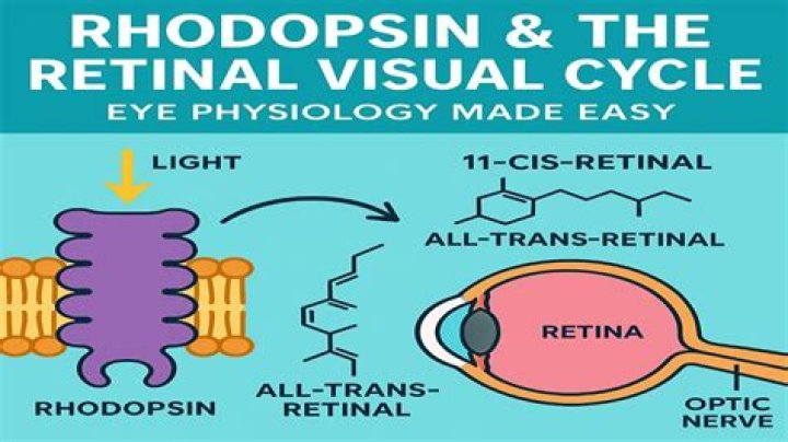
The visual cycle is the biological conversion of a photon into an electrical signal in the retina. This process occurs via G-protein coupled receptors called opsins which contain the chromophore 11-cis retinal. 11-cis retinal is covalently linked to the opsin receptor via Schiff base forming retinylidene protein.
Is retinaldehyde better than retinol?
Retinaldehyde is a rare form of vitamin A that is even more powerful than retinol. As explained earlier, the key difference is that retinaldehyde is much closer in power to retinoic acid, but without the infamous side effects.What type of protein is opsin?
Opsins are membrane proteins with molecular masses of 30-50 kDa that are related to the protein moiety of the photoreceptive molecule rhodopsin; they typically act as light sensors in animals [1-4].
What Happens When rhodopsin is bleached?Bleaching limits the degree to which the rods are stimulated, decreasing their sensitivity to bright light and allowing cone cells (the other type of photoreceptor in the retina) to mediate vision in bright environments.
Article first time published onWhat do melanopsin receptors do?
Melanopsin expressing retinal ganglion cells represent a third class of ocular photoreceptors and are involved in irradiance detection and non-image-forming responses to light including pupil constriction, circadian entrainment, and regulation of sleep.
Does opsin change shape?
The molecule undergoes a series of shape changes to try and better fit the binding site. Therefore, a series of changes in the protein occurs to expel the trans-retinal from the protein.
Is melatonin an opsin?
In vertebrates, both diurnal and nocturnal species exhibit melatonin secretion at night. In mammals, one specific type II opsin, melanopsin (Opn4), links light reception in the eye with melatonin release [8,9].
Does retinal dissociate from opsin?
Retinal rod and cone pigments consist of an apoprotein, opsin, covalently linked to a chromophore, 11-cis retinal. … This dissociation, apparently a general property of cone pigments, results in a surprisingly large amount of free opsin—about 10% of total opsin—in dark-adapted red cones.
Are opsin proteins homologous?
Opsins are operationally defined here as 7-transmembrane proteins structurally and sequentially homologous to GPCR with (Schiff base) lysine in TM7 in alignment with K296 of bovine rhodopsin (or any established opsin).
Is opsin a pigment?
In the cones, the photosensitive pigment is opsin, a transmembrane protein that is very similar to rhodopsin.
What is the purpose of Phototransduction?
Phototransduction is the conversion of light into a change in the electrical potential across the cell membrane. This process involves the sequential activation of a series of signaling proteins, leading to the eventual opening or closing of ion channels in the photoreceptor cell membrane.
How do different organisms sense light and what is the role different opsin proteins have in vision?
Vision depends on detecting light All the visual cells identified in animals detect light using a single family of proteins, called the opsins. … The opsin in turn changes its own shape and turns on signaling pathways in photoreceptor cells that ultimately send a message to the brain that light has been detected.
Which cells in the eye are responsible for scotopic vision?
The retina is comprised of two types of photoreceptor cells: rods and cones. Rods are the cells primarily responsible for scotopic vision, or low-light vision.
What are retinas?
The retina is a layer of tissue in the back of your eye that senses light and sends images to your brain. In the center of this nerve tissue is the macula. It provides the sharp, central vision needed for reading, driving and seeing fine detail. Retinal disorders affect this vital tissue.
Is image formed on blind spot?
In the blind spot, there are no photoreceptors i.e. neither rods or cones, and, therefore, there is no image formation in this area.
What is scintillating scotoma?
Scintillating scotomas are blind spots that flicker and waver between light and dark. Scintillating scotomas are typically not permanent. But they can be an indicator of an underlying health condition.
How does opsin absorb light?
Most animals capture light with opsins for vision and non-visual functions. Opsins form light-sensitive pigments with a retinal chromophore and absorb light to activate G proteins. Therefore, opsins are considered to be light-sensitive G protein coupled receptors (GPCRs).
Is opsin a protein?
Opsins are membrane proteins with molecular masses of 30-50 kDa that are related to the protein moiety of the photoreceptive molecule rhodopsin; they typically act as light sensors in animals [1–4].
Is mouse opsin an ion channel?
Some contain opsins like channelrhodopsin 2 (ChR2, or COP4) from the algae Chlamydomonas reinhardtii. They are sodium ion channels and respond to blue light.
How long does it take for Retinaldehyde to work?
At 18 weeks, retinaldehyde and retinoic acid results showed a significant reduction in wrinkles and roughness. The effect at 44 weeks was still significant but less pronounced.
Is Retinaldehyde irritating?
Retinaldehyde is a stabilized form of Vitamin A that slowly converts to Retinoic Acid in the skin. Retinaldehyde is a more gentle version in the Retinoic family. Like Retinol, Retinaldehyde rejuvenates the skin and improves its cellular-turnover rate, but in a less-irritating formulation.
Can I use Retinaldehyde every day?
Retinaldehyde is non-irritating to the skin and can be used daily AM and PM, unlike many other forms of Vitaimin A.
How do photons affect rhodopsin?
Photon absorption causes bleaching of rhodopsin, a process that is initiated by the photoconversion of 11-cis-retinal to all-trans-retinal. … This interaction initiates a cyclic nucleotide cascade that ultimately converts the absorbed light into an electrical response of the rod cell.
What is the chemical reaction in rhodopsin that is involved in vision?
Retinol can be convert to retinal, and retinal is a chemical necessary for rhodopsin. As light enters the eye, the 11-cis-retinal is isomerized to the all-“trans” form.
How does rhodopsin function as a light receptor?
Rhodopsin is a protein that is essential for vision, especially in dim light. The photoreceptors in the retina that contain rhodopsin are rods. Rhodopsin is attached to 11-cis retinal which becomes excited by a photon of light and isomerizes to become all-trans conformation.