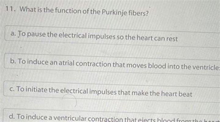
What is the function of the Purkinje fibers
 John Peck
 Purkinje fibers play a major role in electrical conduction and propagation of impulse to the ventricular muscle.
What is the function of Purkinje fibers quizlet?
What is the function of the purkinje fibers? Send nerve impulses to the cells in the ventricles of the heart and cause them contract and pump blood either to the lungs or the rest of the body.
What is heart Purkinje fibers?
The Purkinje fibres (sub-endocardial plexus of conduction cells) are a network of specialised cells. … These cells are located in the subendocardial surface of the ventricular walls, and are able to rapidly transmit cardiac action potentials from the atrioventricular bundle to the myocardium of the ventricles.
Where are the Purkinje fibers located and what is their function?
Purkinje fibers (or Purkyne tissue) are located in the inner ventricular walls of the heart, just beneath the endocardium. These fibers are specialized myocardial fibers that conduct an electrical stimulus or impulse that enables the heart to contract in a coordinated fashion.Where are Purkinje Fibres?
The purkinje fibres are found in the sub-endocardium. They are larger than cardiac muscle cells, but have fewer myofibrils, lots of glycogen and mitochondria, and no T-tubules. These cells are connected together by desmosomes and gap junctions, but not by intercalated discs.
What do the Purkinje fibers connect?
The Purkinje fibers connect with the ends of the bundle branches to form interweaving networks on the endocardial surface of both ventricles and transmit the cardiac impulse almost simultaneously to the entire right and left ventricular endocardium.
What is the role of the Purkinje system in causing the ventricular muscle to synchronously contract?
The His-Purkinje System (HPS) is responsible for the rapid electric conduction in the ventricles. It relays electrical impulses from the atrioventricular node to the muscle cells and, thus, coordinates the contraction of ventricles in order to ensure proper cardiac pump function.
Why do Purkinje fibers conduct the fastest?
The fast propagation is partially due to the different connexins in the gap junctions in these cells. The amount of Cx40, a connexin protein that causes high conductance channels, is at least three fold greater in Purkinje fibers than in myocardial cells.What would happen if the Purkinje Fibres did not do their function?
If the AV node also fails, Purkinje fibers are capable of acting as the pacemaker. … When the electrical activity reaches the atrio-ventricular node it is slowed down- an important function of the AVN that allows the atria to fully contract before the ventricles contract.
What are Purkinje cells?Purkinje cells are a unique type of neuron-specific to the cerebellar cortex. They are remarkable (and instantly recognizable) for their massive, intricately branched, flat dendritic trees, giving them the ability to integrate large amounts of information and learn by remodeling their dendrites.
Article first time published onAre Purkinje fibers muscle cells?
Purkinje fibers are specialized cardiac muscle cells that conduct electrical impulses that allow coordinated contraction of cardiac muscle. In this specimen, the aorta is on the left and the left ventricle on the right. Purkinje fibers are found in the inner ventricular wall beneath the endocardium.
Are Purkinje Fibres nerves?
Purkinje fibres are nerve fibres supplying the ventricular muscle.
What stimulates the Purkinje fibers?
Sympathetic stimulation increases the slope of the pacemaker potential and depolarizes the resting membrane potential. Both of these help increase the heart rate. Sympathetic stimulation releases norepinephrine that acts on the SA node through β1 receptors that are coupled to a Gs protein.
What are Purkinje fibers derived from?
Purkinje fibers from dogs and sheep are the most frequently used; however, fibers have also been obtained from other animals such as calves, pigs, goats, and rabbits. The number of usable fibers varies with each heart, and in the dog ranges from three to eight.
What is the purpose of the Endocardium?
Definition and Function Anatomic function: A tissue covering the inside of the heart, the endocardium keeps the blood flowing through the heart separate from the myocardium, or cardiac muscles. It also lines the valves, which open and close to regulate blood flow through the chambers of the heart.
How are Purkinje fibers activated?
In ventricular, atrial, and Purkinje fibers, the action potential begins with a phase ofrapid depolarization, called the upstroke. As in nerve and skeletal muscle, the upstroke is caused by a transient increase in Na+ conductance (gNa), produced by depolarization-induced opening of activation gates on the Na+ channels.
What is his Purkinje network?
The Purkinje network is a specialized conduction system within the heart that ensures the proper activation of the ventricles to produce effective contraction.
What is the bundle of His and Purkinje Fibres?
Bundle of His is a collection of specialized heart muscle cells that transmit electrical impulses from the AV node in the heart to the muscle cells of the heart wall. Meanwhile, Purkinje fibres are thin filaments that distribute electrical impulses to the ventricle myocardium and activate right and left ventricles.
What is the function of bundle of his?
Function. The bundle of His is an important part of the electrical conduction system of the heart, as it transmits impulses from the atrioventricular node, located at the anterior-inferior end of the interatrial septum, to the ventricles of the heart.
How do electrical impulses pass from Purkinje fibers to ventricular cardiac muscle fibers?
Both bundle branches descend and reach the apex of the heart where they connect with the Purkinje fibers (see image above, step 4). This passage takes approximately 25 ms. The Purkinje fibers are additional myocardial conductive fibers that spread the impulse to the myocardial contractile cells in the ventricles.
Why do the Purkinje fibers cause the papillary muscles to contract before the ventricles contract?
a. Junctional fibers are small, allowing the atria to contract before the impulse spreads rapidly over the ventricles. … Branches of the A-V bundle give rise to Purkinje fibers leading to papillary muscles; these fibers stimulate contraction of the papillary muscles at the same time the ventricles contract.
Can Purkinje fibers self excite?
Can Purkinje fibers self-excite? – Quora. It appears so. No, they’re modified cardiac muscle cells that behave like nerve fibers. Their function, unlike that of other cardiac muscle, is rapid electrical conduction of nervelike signals.
Are Purkinje fibers in the myocardium?
Purkinje fibers are specialized conductive myocardial cells with few myofibrils that promotes the rapid conduction of the impulse through the ventricles. They are larger than the normal myocardial fibers. The waves of depolarization ultimately spread to adjacent myocardial cells through the intercalated discs.
What layer of the heart are Purkinje fibers found?
Purkinje fibres lie in the deepest layer of the endocardium and supply the papillary muscles.
Are Purkinje fibers multipolar?
The Purkinje cell, a multipolar neuron in the cerebellum, has many branching dendrites, but only one axon. Pseudounipolar cells share characteristics with both unipolar and bipolar cells.
What are the cerebellum's functions?
The cerebellum is important for making postural adjustments in order to maintain balance. Through its input from vestibular receptors and proprioceptors, it modulates commands to motor neurons to compensate for shifts in body position or changes in load upon muscles.
Are Purkinje tissue modified myocardial cells?
Purkinje fibers are modified cardiac muscle cells and stain differently from surrounding muscle cells: Larger cells. Large amounts of glycogen. Fewer myofibrils.
Where is the Purkinje fibers located quizlet?
purkinje fibers- located in the walls of the ventricles.