What is a Conus
 Christopher Lucas
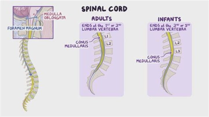
 The conus medullaris (Latin for “medullary cone”) or conus terminalis is the tapered, lower end of the spinal cord. It occurs near lumbar vertebral levels 1 (L1) and 2 (L2), occasionally lower. … The filum terminale provides a connection between the conus medullaris and the coccyx which stabilizes the entire spinal cord.
What is the conus of the spine?
Conus medullaris – The cone-shaped bottom of the spinal cord, usually at the level of L1. Disc (Intervertebral) – A tough, elastic cushion located between the vertebrae in the spinal column; acts as a shock absorber for the vertebrae.
What level is conus?
The conus medullaris is the terminal end of the spinal cord, which typically occurs at the L1 vertebral level in the average adult.
What are conus ends?
As mentioned above, the conus medullaris is the distal tapering end of the spinal cord. … On average, the conus terminates at the middle third of the L1 vertebra but can be located as high as the middle third of the T11 vertebra or as low as the middle third of L3 vertebra.What is normal conus?
The range of conus levels for the entire group of normal children was T12 to L3. … A conus level at L2-L3 or above should be considered normal at any age. A conus level at L3 is indeterminate, since it is possible for a normal or a tethered conus to be located at this level.
What part of your spine controls your legs?
The nerves in your thoracic spine go to your chest and abdomen. The nerves of the lumbar spine then reach to your legs, bowel, and bladder. These nerves coordinate and control all the body’s organs and parts, and let you control your muscles.
What is the cauda?
Cauda is Latin for tail, and equina is Latin for horse (ie, the “horse’s tail”). The CE provides sensory innervation to the saddle area, motor innervation to the sphincters, and parasympathetic innervation to the bladder and lower bowel (ie, from the left splenic flexure to the rectum).
What are the first signs of cauda equina?
- Lower limb weakness and intermittent changes in sensation, such as numbness.
- “Saddle anesthesia” – loss or diminished sensation in areas where a person would sit on a saddle.
- Urinary and/or bowel problems, such as retention or incontinence.
Where does Conus normally terminate?
Conclusions: The CM terminates most commonly at the L1-2 disc space and in the absence of tethering, the CM virtually never ends below the mid-body of L2. A CM that appears more caudal on neuroimages should be considered tethered.
Where is the epidural space?The epidural space is the area between the dura mater (a membrane) and the vertebral wall, containing fat and small blood vessels. The space is located just outside the dural sac which surrounds the nerve roots and is filled with cerebrospinal fluid.
Article first time published onWhat is a low lying Conus?
Low-lying conus medullaris: It refers to a low position of a normal-appearing conus medullaris with respect to the vertebral level. It is usually located between the T12–L1 and L1–L2 disk level; however, in 6.4% of population it can be found between the upper and middle third of L2.
Why is cauda equina an emergency?
When the Cauda Equina nerves are compressed this normally results in what are commonly referred to as ‘red flag’ symptoms. Cauda Equina Syndrome is a medical emergency because delayed decompression surgery can result in lifelong disability.
What is the Conus equina?
The collection of nerves at the end of the spinal cord is known as the cauda equina, due to its resemblance to a horse’s tail. The spinal cord ends at the upper portion of the lumbar (lower back) spine.
Is lumbar spondylosis arthritis?
Technically, spondylosis is a form of arthritis—spinal osteoarthritis (osteoarthritis is the most common type of arthritis) to be exact. We tend to think of arthritis as something you get in your hands and knees, but the spine, and all of its bones and joints, can fall victim to its grip as well.
What is Foraminal narrowing in lumbar spine?
Foraminal narrowing, or foraminal stenosis, is a condition of the spine that can cause pain and other symptoms resulting from spinal nerve root compression. At every level of the spine, a pair of nerve roots runs through the spinal column via small openings called foramina (singular: foramen).
Is spondylolisthesis a disease?
Spondylolisthesis is a spinal condition that affects the lower vertebrae (spinal bones). This disease causes one of the lower vertebrae to slip forward onto the bone directly beneath it. It’s a painful condition but treatable in most cases.
Is neurogenic claudication an emergency?
It is therefore considered a medical emergency. Treatment for spinal stenosis consists of surgery to relieve the pressure on the spinal cord or nerve roots.
What does no reflexes in legs mean?
When reflex responses are absent this could be a clue that the spinal cord, nerve root, peripheral nerve, or muscle has been damaged. When reflex response is abnormal, it may be due to the disruption of the sensory (feeling) or motor (movement) nerves or both.
What's the cause of sciatica?
Sciatica most commonly occurs when a herniated disk, bone spur on the spine or narrowing of the spine (spinal stenosis) compresses part of the nerve. This causes inflammation, pain and often some numbness in the affected leg.
Which bones protect the brain?
The skull protects the brain and forms the shape of the face. The spinal cord, a pathway for messages between the brain and the body, is protected by the backbone, or spinal column.
What is the best exercise for the spine?
- Knee-to-chest stretch. Knee-to-chest stretches elongate your spine and reduce lower back pain. …
- Rotational stretch. …
- Pelvic tilt. …
- Bridge. …
- Partial abdominal curl. …
- Cat-cow stretch. …
- Shoulder blade squeeze. …
- Chin-to-chest stretch.
What part of the spine controls the heart?
Thoracic (mid back) – the main function of the thoracic spine is to hold the rib cage and protect the heart and lungs. The twelve thoracic vertebrae are numbered T1 to T12.
What levels cause cauda equina syndrome?
Cauda equina syndrome (CES) results from compression and disruption of the function of these nerves and can be inclusive of the conus medullaris or distal to it, and most often occurs when damage occurs to the L3-L5 nerve roots.
What is mild neural Foraminal stenosis?
Neural foraminal stenosis refers to the narrowing of the small openings between each vertebra in the spine, called foramen, which nerve roots pass through. A type of spinal stenosis, neural foraminal stenosis, does not always cause symptoms. But if a nerve gets compressed in the gap, this will be painful.
What is the difference between cauda equina and spinal cord compression?
Spinal cord compression and Cauda Equina Syndrome have similar symptoms, including back pain and weakness or paralysis of the lower limbs. This means the relatively rare Cauda Equina Syndrome is often misdiagnosed as spinal cord compression, resulting in the right treatment often not being given in time.
What is the most common cause of cauda equina syndrome?
- A severe ruptured disk in the lumbar area (the most common cause)
- Narrowing of the spinal canal (stenosis)
- A spinal lesion or malignant tumor.
- A spinal infection, inflammation, hemorrhage, or fracture.
How serious is cauda equina syndrome?
Cauda equina syndrome is a rare but serious condition that describes extreme pressure and swelling of the nerves at the end of the spinal cord. Cauda equina syndrome is a medical emergency that calls for urgent surgical intervention.
Can you fully recover from cauda equina syndrome?
Although cauda equina syndrome is not a fatal condition, it can cause severe neurological damage. If the condition is not treated quickly enough, this damage may be irreversible, meaning a patient will not make a full recovery.
What is the subdural space?
The subdural space is a potential intracranial space situated between the arachnoid and dura. Fluid can collect in the subdural space and in the subarachnoid space. … Extraaxial fluid collections are found in 20% to 50% of children with bacterial meningitis who have cranial CT performed.
Is cerebrospinal fluid found in the subdural space?
The classic view has been that a so-called subdural space is located between the arachnoid and dura and that subdural hematomas or hygromas are the result of blood or cerebrospinal fluid accumulating in this (preexisting) space.
Does the brain have an epidural space?
In anatomy, the epidural space is the potential space between the two layers of the dura mater (the outermost meningeal layer that covers the brain and spinal cord).