What causes P waves
 Andrew Campbell
 The P wave indicates atrial depolarization. The P wave occurs when the sinus node, also known as the sinoatrial node, creates an action potential that depolarizes the atria.
What do P waves indicate?
The P wave represents the electrical depolarization of the atria. In a healthy person, this originates at the sinoatrial node (SA node) and disperses into both left and right atria.
What happens during the P wave?
The P wave represents the depolarization of the left and right atrium and also corresponds to atrial contraction. Strictly speaking, the atria contract a split second after the P wave begins. Because it is so small, atrial repolarization is usually not visible on ECG.
What causes P wave elevation?
The Abnormal P wave Elevation or depression of the PTa segment (the part between the p wave and the beginning of the QRS complex) can result from atrial infarction or pericarditis. If the p-wave is enlarged, the atria are enlarged.What causes a P wave the QRS complex and at wave?
Atrial and ventricular depolarization and repolarization are represented on the ECG as a series of waves: the P wave followed by the QRS complex and the T wave. The first deflection is the P wave associated with right and left atrial depolarization. Wave of atrial repolarization is invisible because of low amplitude.
How does the P wave relate to heart function?
The first wave (p wave) represents atrial depolarisation. When the valves between the atria and ventricles open, 70% of the blood in the atria falls through with the aid of gravity, but mainly due to suction caused by the ventricles as they expand.
What is normal P wave in ECG?
P-wave checklist The P-wave is frequently biphasic in V1 (occasionally in V2). The negative deflection is normally <1 mm. P-wave duration should be ≤0,12 seconds. P-wave amplitude should be <2,5 mm in the limb leads.
What is an abnormal P axis?
Abnormal P-wave axis is defined as any value outside 0–75° (Figure 1) (31). Figure 1. Representative ECG tracings of abnormal P-wave indices. A through (D), Prolonged P-wave duration (A), abnormal P-wave axis (B), abnormal P-wave terminal force in V1 (C), and advanced interatrial block (D).Where do P waves originate?
The P wave is a summation wave generated by the depolarization front as it transits the atria. Normally the right atrium depolarizes slightly earlier than left atrium since the depolarization wave originates in the sinoatrial node, in the high right atrium and then travels to and through the left atrium.
Why is there no P wave in atrial fibrillation?Because the atrial rate is so fast, and the action potentials produced are of such low amplitude, P waves will not be seen on the ECG in patients with atrial fibrillation.
Article first time published onWhat do you see in P waves before QRS?
P waves are the key to determining whether a patient is in sinus rhythm or not. If P waves are not clearly visible in the chest leads, look for them in the other leads. The presence of P waves immediately before every QRS complex indicates sinus rhythm.
How do you find P waves?
Finding the Distance to the Epicenter (From Bolt, 1978.) Measure the distance between the first P wave and the first S wave. In this case, the first P and S waves are 24 seconds apart. Find the point for 24 seconds on the left side of the chart of simplified S and P travel time curves and mark that point.
What is the difference between P wave and T wave?
P-WaveT-WaveA P-wave is produced due to the depolarization of the atrial musculatureA T-wave is produced due to the repolarization of ventricular musculature.
What does P QRS and T wave represent?
The P wave in an ECG complex indicates atrial depolarization. The QRS is responsible for ventricular depolarization and the T wave is ventricular repolarization.
Should I worry about abnormal ECG?
Most of the time severe abnormalities that pop up without any other symptoms are a sign of improper lead placement or an incorrect ECG procedure. However, markedly abnormal ECGs with symptoms are considered a medical emergency that requires treatment or surgery.
What does a prolonged P wave mean?
Prolonged P wave duration signifies conduction delay between right and left atrium due to impulse slowing or blockage, probably most often but not exclusively in the Bachmann bundle. On the ECG this conduction delay is referred to as interatrial block (IAB) [14–16].
In which lead is the P wave best seen?
Sinus P waves are usually most prominently seen in leads II and V1. A negative P wave in lead I may be due to incorrect recording of the electrocardiogram (that is, with transposition of the left and right arm electrodes), dextrocardia, or abnormal atrial rhythms. The P wave in V1 is often biphasic.
What is the name of the body's natural pacemaker?
The sinus node is sometimes called the heart’s “natural pacemaker.” Each time the sinus node generates a new electrical impulse; that impulse spreads out through the heart’s upper chambers, called the right atrium and the left atrium (figure 2).
What heart rhythm has no P wave?
A junctional rhythm is characterized by QRS complexes of morphology identical to that of sinus rhythm without preceding P waves.
Can You Feel P waves?
The waves also travel through the Earth at different speeds. The fastest wave, called the “P” (primary) wave, arrives first and it usually registers a sharp jolt. … “It feels more abrupt, but it attenuates very quickly, so if you are far away you often won’t feel the P wave.”
What waves cause earthquakes?
Seismic waves are caused by the sudden movement of materials within the Earth, such as slip along a fault during an earthquake. Volcanic eruptions, explosions, landslides, avalanches, and even rushing rivers can also cause seismic waves.
What is a terminal P-wave?
The terminal portion of the P-wave in the surface ECG mostly represents the activation of the left atrium. 20. Increases in the negative terminal portion of the P-wave in lead V1 have been reported to be linked to left atrial hypertrophy detected by autopsy21 and LAE detected by echocardiography.
Does atrial fibrillation show up on echocardiogram?
If you have atrial fibrillation, a number of other tests may be carried out, including: an echocardiogram – an ultrasound scan of the heart, which can help identify any other heart-related problems; it’s used to assess the structure and function of the heart and valves.
Where does a rhythm without P waves originate?
A junctional rhythm is an abnormal heart rhythm that originates from the AV node or His bundle.
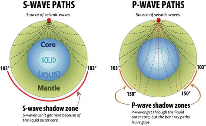
What type of body wave Cannot travel through the liquid layer of the earth?
S-waves cannot travel through liquids. When they reach the surface they cause horizontal shaking. Liquids don’t have any shear strength and so a shear wave cannot propagate through a liquid.
What is the difference between the arrival time of P and S waves?
S waves are indicated by an abrupt change in wave amplitude. In the seismogram below, we see that the S wave arrived at about 34 sec after the P wave arrived. This time difference is called the S-P interval and is the lag time between the P and S wave.
How do you calculate P wave on ECG?
The best way to determine the ventricular heart rate would be to simply count the QRS complexes and multiply by 6, which would be 15 x 6 = 90 bpm. The P waves are not able to be identified in atrial fibrillation, and it is assumed that the atrial rate is between 400 and 600 bpm.
What does the QRS stand for?
Answer. The QRS duration represents the time for ventricular depolarization. The duration is normally 0.06 to 0.10 seconds. Q waves are inscribed when the initial QRS vector is directed away from the positive electrode.
What does the P wave represent Class 11?
P wave: It is the first wave of ECG of duration 0.1 sec, directed upwards, rounded or pointed. It is due to atrial depolarization and also represents the spread of impulse from SA Node to atrial muscles. Its height is up to 0.5 mV which represents the functional activity of atrial muscles.
What does T wave mean?
The T wave on an electrocardiogram (ECG) represents typically ventricular repolarization.
Is Sinus Arrhythmia serious?
Keep in mind that for the majority of people, a sinus arrhythmia is neither dangerous nor problematic. Even if your doctor suspects you have this irregular heartbeat, he may not order the test to check for it. That’s because an EKG can be costly, and a sinus arrhythmia is considered a benign condition.