What are the meninges composed of
 Andrew White
 The meninges are three protective membrane layers surrounding the brain and spinal cord. They are composed of the pia (closest to the CNS), arachnoid, and dura (outermost layer), and contain blood vessels and enclose the cerebrospinal fluid.
What 3 layers make up the meninges?
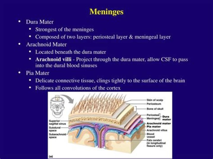
Three layers of membranes known as meninges protect the brain and spinal cord. The delicate inner layer is the pia mater. The middle layer is the arachnoid, a web-like structure filled with fluid that cushions the brain. The tough outer layer is called the dura mater.
What are meningeal membranes made of?
Meninges are formed by three tissue membranes that are primarily known as wrappers of the brain. They consist of dura mater, arachnoid and pia mater. The dura mater or pachymeninx (pachy-thick) is the outer membrane and forms a sac that envelops the other meningeal layers.
What cells are in meninges?
The principal cell types in meninges are fibroblasts within the stroma and the vascular endothelium. The extent of collagen production is greater in the thicker outer dura mater and relatively less in the two thinner leptomeningeal layers, the arachnoid and pia mater.What are meninges filled with?
The middle layer of meninges is arachnoid, a thin layer resembling a cobweb with numerous threadlike strands attaching it to the innermost layer. … The space under the arachnoid, the subarachnoid space, is filled with cerebrospinal fluid and contains blood vessels.
What are the three meninges quizlet?
there are three meninges: the dura mater, arachnoid membrane/layer, and the pia mater.
What is the arachnoid villi?
Arachnoid granulations or villi are growths of arachnoid membrane into the dural sinuses, through which the CSF enters the venous system from the subarachnoid space. 1. Arachnoid villi are microscopic, whereas arachnoid granulations represent distended villi and are visible to the naked eye.
How are meninges linked to each other?
The two dural layers are firmly attached to each other, except in places where they separate to enclose the dural venous sinuses. In these places, the meningeal layer projects inward, towards the cerebral tissue, forming the fibrous septa that partially separate the cranial cavity.What is the arachnoid mater made of?
The arachnoid is composed of collagen and elastic fibers. It has a variable thickness, in places being formed by several cell layers. Its outer (dural) aspect is smoother than the inner (pial) aspect from which trabeculae emerge to bridge the subarachnoid space (Nicholas and Weller, 1988).
What is the most superficial layer of the meninges?The most superficial layer of the meninges is the dura mater.
Article first time published onWhat is meninges in biology?
The meninges are membranous layers surrounding the central nervous system. In the head, the meninges lie between the brain and the skull, and interact closely with both during development.
What are the meninges quizlet?
Protect the brain from injury and is made up of three layers: dura mater, arachnoid mater, and pia mater. … Dura mater. Two layers: superficial Periosteal Layer attachs to the skull and deeper Meningeal Layer forms the true external covering of the brain.
Which meninges are vascular?
The blood supply of the meninges generally concerns the blood supply of the outer layer of dura mater rather than the inner layer of dura mater, arachnoid or pia mater which do not require a large blood supply. There are several arteries that supply the dura in the anterior, middle, and posterior cranial fossae 1,2.
Does pia mater contain CSF?
Function. In conjunction with the other meningeal membranes, pia mater functions to cover and protect the central nervous system (CNS), to protect the blood vessels and enclose the venous sinuses near the CNS, to contain the cerebrospinal fluid (CSF) and to form partitions with the skull.
What is spiral cord?
A column of nerve tissue that runs from the base of the skull down the center of the back. It is covered by three thin layers of protective tissue called membranes. The spinal cord and membranes are surrounded by the vertebrae (back bones).
What are the arachnoid villi and its function?
Physiology of the arachnoid villi Arachnoid villi act as one-way valves for the flow of CSF into venous blood, and hydrostatic pressure is the main stimulus that causes these valves to open.
What are the three main regions of the brainstem?
The brainstem is divided into three sections in humans: the midbrain (mesencephalon), the pons (metencephalon), and the medulla oblongata (myelencephalon).
What are the 3 meninges and where are they found?
meninges, singular meninx, three membranous envelopes—pia mater, arachnoid, and dura mater—that surround the brain and spinal cord. Cerebrospinal fluid fills the ventricles of the brain and the space between the pia mater and the arachnoid.
What layers make up the meninges quizlet?
The three layers of the meninges are the dura mater, arachnoid, and pia mater.
What are the different parts of the central nervous system?
The central nervous system (CNS) controls most functions of the body and mind. It consists of two parts: the brain and the spinal cord. The brain is the center of our thoughts, the interpreter of our external environment, and the origin of control over body movement.
What type of tissue is pia mater?
The term “pia mater” means “tender matter.” It is composed of delicate connective tissue and has many tiny blood vessels. The pia mater is the only layer that clings tightly to the brain and follows all of its convolutions. Cerebral arteries and veins travel in the subarachnoid space, completely enveloped by pia mater.
What type of tissue is the arachnoid?
structure of meninges …the subarachnoid space is the arachnoid, a thin, transparent membrane. It is composed of fibrous tissue and, like the pia mater, is covered by flat cells also thought to be impermeable to fluid.
Is the dura mater vascular?
Blood Supply and Lymphatics The dura mater receives vascular supply from the following branches: Internal carotid artery. Maxillary artery.
What layer of the meninges is closest to the brain?
Dura Mater The outermost mater of the meninges, the dura, is composed of two layers: the periosteal layer that lies closest to the calvarium and the meningeal layer that lies closest to the brain tissue. These together contribute to the dura being a thick, dense, fibrous membrane that is quite inelastic.
What structure forms choroid plexus?
The choroid plexus, or plica choroidea, is a plexus of cells that arises from the tela choroidea in each of the ventricles of the brain. The choroid plexus produces most of the cerebrospinal fluid (CSF) of the central nervous system.
What structure in the ventricles produces CSF?
The choroid plexuses are located in the ventricles produce CSF, which fills the ventricles and subarachnoid space, following a cycle of constant production and reabsorption.
Which type of cell is composed of one dendrite and one axon and is found in the eye and nose *?
A bipolar neuron has one axon and one dendrite extending from the soma. An example of a bipolar neuron is a retinal bipolar cell, which receives signals from photoreceptor cells that are sensitive to light and transmits these signals to ganglion cells that carry the signal to the brain.
What is the space between a dendrite and an axon called?
The space between the dendrites of one neuron and the axon of another neuron is called the synapse.
What is the midbrain?
The midbrain is the topmost part of the brainstem, the connection central between the brain and the spinal cord. There are three main parts of the midbrain – the colliculi, the tegmentum, and the cerebral peduncles.
Where is the origin of the meninges?
The cranial meninges originate from a mesenchymal sheath on the surface of the developing brain, called primary meninx, and undergo differentiation into three layers with distinct histological characteristics: the dura mater, the arachnoid mater, and the pia mater.
What is CNS in medical terms?
The Central Nervous System (CNS) includes the brain and spinal cord, while Peripheral Nervous System (PNS) includes nerves connected to the spinal cord.