What are the cords of the brachial plexus
 Isabella Bartlett
 RootsC5, C6, C7, C8, T1CordsLateral Medial PosteriorTerminal branchesMusculocutaneous nerve Axillary nerve Radial nerve Median nerve Ulnar nerveInnervationComplete sensory and motor innervation of the arm
How many cords does the brachial plexus have?
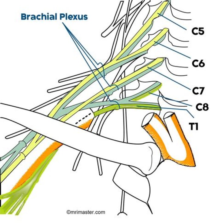
Brachial plexus architecture Typically, the brachial plexus is composed of 5 roots, 3 trunks, 6 divisions, 3 cords, and terminal branches, as seen in the image below. Brachial plexus with terminal branches labeled. MC is musculocutaneous (nerve), AXI is axillary, RAD is radial, MED is median, and ULN is ulnar.
What are the branches of lateral cord?
Schematic Anatomy. The terminal branches of the lateral cord are the lateral pectoral nerve and the musculocutaneous nerve (which itself terminates as the lateral cutaneous nerve of the forearm). The lateral cord also contributes to the median nerve (Figure 40.1).
Where does the brachial plexus start and end?
The brachial plexus is a network of nerve fibres that supplies the skin and musculature of the upper limb. It begins in the root of the neck, passes through the axilla, and runs through the entire upper extremity.What is spiral cord?
A column of nerve tissue that runs from the base of the skull down the center of the back. It is covered by three thin layers of protective tissue called membranes. The spinal cord and membranes are surrounded by the vertebrae (back bones).
What is the lumbosacral plexus?
The lumbosacral plexus is a network of nerves derived from lumbar and sacral roots with each one of them dividing into anterior and posterior branches. … The anterior branches supply the flexor muscles of thigh and leg and posterior branches supply the extensor and abductor muscles.
What are the roots trunks divisions and cords of the brachial plexus?
RootsC5, C6, C7, C8, T1CordsLateral Medial PosteriorTerminal branchesMusculocutaneous nerve Axillary nerve Radial nerve Median nerve Ulnar nerveInnervationComplete sensory and motor innervation of the arm
What is the lateral cord?
The lateral cord is the part of the brachial plexus formed by the anterior divisions of the upper (C5-C6) and middle trunks (C7). Its name comes from it being lateral to the axillary artery as it passes through the axilla.Is the ulnar nerve Part of the brachial plexus?
Ulnar nerve The C8 and T1 roots are part of the brachial plexus that travels from the cervical spine, under the clavicle, through the armpit (axilla), and down the inside of the arm to the inner elbow.
What muscles are innervated by the posterior cord of the brachial plexus?NameRootsSuppliesradial nerveC5-C8, T1triceps brachii muscle, the brachioradialis muscle, the extensor muscles of the fingers and wrist (extensor carpi radialis muscle), supinator, and the extensor and abductor muscles of the thumb
Article first time published onWhere is the brachial vein?
The brachial veins are usually 2 in number and they are located on either side of the brachial artery. They are usually formed by the union of the radial and the ulnar venae comitantes, near the level of the elbow [1].
What is the brachial region?
the brachial region encompassing the upper arm, the olecranal region encompassing the back of the elbow, the antebrachial region encompasses the forearm, front and back.
What is the brachial distal third?
It is the smallest and most medial branch of the brachial plexus, originating from C8 and T1 nerve roots. [1] As it descends the arm, it courses with the basilic vein, terminating at the distal third of the medial arm.
What does the brachial plexus innervate?
The brachial plexus is a major network of nerves transmitting signals responsible for motor and sensory innervation of the upper extremities, including the shoulder, arm, and hand.
Which of the following nerves is part of the brachial plexus?
The brachial plexus transmits both motor and sensory information from and to spinal nerves C5, C6, C7, C8, & T1.
What is the cauda?
Cauda is Latin for tail, and equina is Latin for horse (ie, the “horse’s tail”). The CE provides sensory innervation to the saddle area, motor innervation to the sphincters, and parasympathetic innervation to the bladder and lower bowel (ie, from the left splenic flexure to the rectum).
Why does L2 end spinal cord?
It is these spinal nerve roots that compose the cauda equina beyond L1/L2. The fact that the spinal cord ends at L1/L2 is very useful in clinical practice in that it allows for spinal taps to be performed to sample CSF without the risk of puncturing the spinal cord.
Is nerve cord and spinal cord same?
No. Both nerve cord and spinal cord are different from each other. The nerve cord is a hollow tube of nervous tissue and is an important structure of the central nervous system.
What nerve Innervates deltoid?
After exiting the quadrangular space posteriorly, the anterior branch of the axillary nerve wraps around the surgical neck of the humerus, with the posterior humeral circumflex artery, to then innervate the deltoid muscle.
What are the ventral Rami?
The ventral ramus (pl. rami) (Latin for branch) is the anterior division of a spinal nerve. The ventral rami supply the antero-lateral parts of the trunk and the limbs. … In regions other than the thoracic, ventral rami converge with each other to form networks of nerves called nerve plexuses.
Which of the following terminal nerves arises from the lateral cord of the brachial plexus quizlet?
–The medial pectoral nerve arises from the medial or lateral cords of the brachial plexus.
What forms the lumbosacral plexus?
The lumbosacral plexus is formed by the anterior rami of the nerves (spinal segments T12–S4) to supply the lower limbs. The lumbosacral plexus can be divided into the lumbar plexus, which innervates the ventral upper half, and the sacral plexus, which mainly innervates the dorsal side.
Which spinal cord plexus supplies the abdominal wall?
The ventral rami of L1-L5 spinal nerves with a contribution of T12 form Lumbar plexus. This plexus lies within the psoas major muscle. Nervi of the plexus serve the skin and the muscles of the lower abdominal wall, the thigh and external genitals.
Is femoral anterior or posterior?
Femoral nerve is the main nerve of anterior compartment of thigh. It originates from the dorsal sections of the anterior primary rami of L2, L3, L4 nerves and is the largest branch of lumbar plexus.
What connects ulnar nerve?
The ulnar nerve starts at the brachial plexus in the armpit and: Connects to the C8 cervical vertebra and the T1 thoracic vertebra (the middle of the brachial plexus). Runs down the front of the upper arm near the axillary and brachial arteries.
What does the dorsal scapular nerve innervate?
The DSN is a motor nerve that innervates the levator scapulae, rhomboid major, and rhomboid minor muscles. These muscles work dynamically and collectively are considered periscapular stabilizing muscles. Individually they can retract and elevate the scapula.
What nerve innervates the posterior deltoid?
The posterior branch of the axillary nerve innervates the teres minor and also the deltoid. The posterior branch then winds round the deltoid muscle and goes to innervate an area of skin on the back of the arm as the upper lateral brachial cutaneous nerve, or the superior lateral cutaneous nerve of arm.
What is musculocutaneous nerve?
The musculocutaneous nerve innervates the three muscles of the anterior compartment of the arm: the coracobrachialis, biceps brachii, and brachialis. It is also responsible for cutaneous innervation of the lateral forearm.
Where does the dorsal scapular nerve come from?
The dorsal scapular nerve originates from the fifth cervical spinal nerve (ventral ramus) in the majority (75%) of cases, within the posterior cervical triangle deep to the prevertebral fascia. However, this nerve may also receive some contributions from C4 to T1.
What does Thoracodorsal nerve supply?
The thoracodorsal nerve is a pure motor nerve that innervates the latissimus dorsi muscle. … The primary blood supply of the latissimus dorsi muscle is the thoracodorsal artery. The dorsal divisions of T6 to T12 provide the sensory innervation of the skin of the latissimus dorsi muscle.
What makes up posterior cord?
Posterior cord was formed by union of posterior division of C5 and C6 roots with posterior division of middle and lower trunk (there was no upper trunk) in 16.2% of upper extremities. Posterior cord of brachial plexus was present lateral to the second part of axillary artery in 18.9% of upper extremities.