Is facial droop ipsilateral or contralateral
 Emily Sparks
 Facial palsy has rarely been observed even in medullary infarction. However, central-type facial palsy is usually found contralaterally to the infarct area at the level of the rostral medulla. In the present report, we discuss the pathogenesis of the neurological manifestations in a 57-year-old man with hypertension.
Is facial droop ipsilateral or contralateral in stroke?
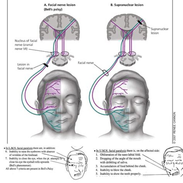
Lesions that damage the motor cortex, such as acute ischemic strokes, will result in contralateral facial weakness of the lower face only, with preservation of the muscles of the upper face on both sides, due to the dual innervation of the upper face.
Which side of the face droops in a stroke?
F.A.S.T. Face drooping is one of the most common signs of a stroke. One side of the face may become numb or weak. This symptom may be more noticeable when the patient smiles. A lopsided grin could indicate that the muscles on one side of the face have been affected.
Is the facial nerve ipsilateral or contralateral?
The left and right branches supply their respective sides of the face (ipsilateral innervation). Accordingly, the posterior components receive motor input from both hemispheres of the cerebral cortex (bilaterally), whereas the anterior components receive strictly contralateral input.Is facial nerve contralateral?
The dorsal aspect of the facial nucleus receives input from both the left and right cerebral hemispheres. This results in both hemispheres having control over the muscles of the upper face. The ventral aspect of the facial nucleus receives mainly contralateral inputs.
What is ipsilateral stroke?
In conclusion, ipsilateral hemiparesis can develop as a result of a new stroke after a previous stroke on the opposite side. The mechanism involved is thought to be functional reorganization of the ipsilateral hemisphere.
What is facial droop?
Facial droop occurs when there is damage to the nerves in the face, preventing the facial muscles from working properly. The nerve damage can either be temporary or permanent. Facial droop can also be caused by damage to the part of the brain that sends nerve signals to the facial muscles.
Where does facial nerve cross?
From the brain stem, the motor and sensory parts of the facial nerve join together and traverse the posterior cranial fossa before entering the petrous temporal bone via the internal auditory meatus.What are the branches of facial nerve?
The facial nerve has five main branches, although the anatomy can vary somewhat between individuals. The branches are, from top to bottom: frontal (or temporal), zygomatic, buccal, marginal mandibular, and cervical. Each of these branches provides input to a group of muscles of facial expression.
Are cranial nerve lesions ipsilateral or contralateral?In summary, all of the cranial nerves lateralized, the ones that don’t cross, are all ipsilateral-ipsilesional. All the ones that cross are the superior rectus subnucleus, nucleus of four, and the upper motor neuron of seven.
Article first time published onWhy does the face droop in a stroke?
Facial paralysis occurs during a stroke when nerves that control the muscles in the face are damaged in the brain. Depending on the type of stroke, damage to the brain cells is caused by either lack of oxygen or excess pressure on the brain cells caused by bleeding.
Which side of the face is affected by a stroke?
The lower part of one side of the face is normally affected (the forehead is usually spared). However, the eye can be involved if the stroke is in the brainstem as the person will experience damage to the facial nucleus; which will present without forehead sparing.
Why would your face droop on one side?
Bell’s palsy, also known as acute peripheral facial palsy of unknown cause, can occur at any age. The exact cause is unknown. It’s believed to be the result of swelling and inflammation of the nerve that controls the muscles on one side of your face. Or it might be a reaction that occurs after a viral infection.
Is facial nerve sympathetic or parasympathetic?
The facial nerve is the seventh cranial nerve. It contains the motor, sensory, and parasympathetic (secretomotor) nerve fibers, which provide innervation to many areas of the head and neck region. The facial nerve is comprised of three nuclei: The main motor nucleus.
Are facial nerves peripheral or central?
The cranial nerves are considered components of the peripheral nervous system (PNS), although on a structural level the olfactory (I), optic (II), and trigeminal (V) nerves are more accurately considered part of the central nervous system (CNS).
What causes corners of mouth to droop?
The main reason for a downturned mouth is the natural ageing process, which can happen to anyone in his or her lifetime. The loss of volume in the cheeks and lateral sides of the face can also cause these lines to appear on the sides of the mouth. Some of the other causes are: Smoking habits.
What is ipsilateral and contralateral?
Contralateral is defined as ‘pertaining to the other side’. Ipsilateral is considered the opposite of contralateral and occurs on the same side.
Is contralateral and ipsilateral the same?
Contralateral: Of or pertaining to the other side. The opposite of ipsilateral (the same side). For example, a stroke involving the right side of the brain may cause contralateral paralysis of the left leg.
What do you mean by ipsilateral?
Listen to pronunciation. (IP-sih-LA-teh-rul) On the same side of the body as another structure or a given point.
What are the names of the facial nerves?
- I. Olfactory nerve.
- II. Optic nerve.
- III. Oculomotor nerve.
- IV. Trochlear nerve.
- V. Trigeminal nerve.
- VI. Abducens nerve.
- VII. Facial nerve.
- VIII. Vestibulocochlear nerve.
Which of the following nerve is a mixed nerve?
The mixed cranial nerves are CN III Occulomotor, CN V Trigeminal, CN VII Facial, CN IX Glossopharyngeal and CN X Vagus.
Where is the chorda tympani located?
Anatomical terms of neuroanatomy The chorda tympani is a branch of the facial nerve that originates from the taste buds in the front of the tongue, runs through the middle ear, and carries taste messages to the brain.
What does the buccal branch of the facial nerve innervate?
The buccal branch of the facial nerve innervates the buccinator, levator labii, anguli oris, and orbicularis oris. Resection margins can also affect drainage of saliva from Stensen’s duct, accessory ducts, and the parotid gland, which may create salivary leaks, fistulae, and sialoceles.
Where are the facial muscles?
- Buccolabial muscles in and around your mouth.
- Nasal muscles around your nose.
- Epicranial muscles of your forehead, skull and neck.
- Auricular muscles around your ears.
- Orbital muscles surrounding your eyes.
Is trigeminal nerve ipsilateral?
Some sensory information from the teeth and jaws is sent from the principal nucleus to the ipsilateral thalamus via the small dorsal trigeminal tract. Touch-position information from the teeth and jaws of one side of the face is represented bilaterally in the thalamus and cortex.
Is Glossopharyngeal nerve ipsilateral?
Glossopharyngeal (CN9): ipsilateral loss of pharyngeal sensation.
Are cranial nerve palsies ipsilateral?
The abducens nerve, also known as cranial nerve VI, is responsible for ipsilateral eye abduction. Visualizing the anatomy of the nerve allows for better appreciation of the causes of abducens nerve palsy.
Can you have a stroke without your face drooping?
Many people are aware of the obvious signs of a stroke such as an excess drooping of the face due to relaxed muscles, but the fact is, there can also be silent stroke symptoms. This means it is completely possible to have a stroke without even noticing.
When a stroke affects the left side?
The effects of a left hemisphere stroke may include: Right-sided weakness or paralysis and sensory impairment. Problems with speech and understanding language (aphasia) Visual problems, including the inability to see the right visual field of each eye.
Which side is worse for stroke?
The terms Left Brain Stroke and Right Brain Stroke refer to the side of the brain where the obstruction causing the stroke occurs. There is not a worse or better side to have a stroke on as both sides control many important functions, but a more severe stroke will result in amplified effects.
What is the difference between hemorrhagic stroke and ischemic stroke?
An ischemic stroke is when blood vessels to the brain become clogged. A hemorrhagic stroke is when bleeding interferes with the brain’s ability to function.