How many fetal shunts are there
 John Peck
 The fetal circulatory system
What are the 3 fetal shunts?
- Ductus arteriosus. � protects lungs against circulatory overload. � allows the right ventricle to strengthen. …
- Ductus venosus. � fetal blood vessel connecting the umbilical vein to the IVC. …
- Foramen ovale. � shunts highly oxygenated blood from right atrium to left atrium.
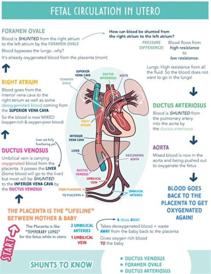
What are the three shunts in the fetal circulation which provide the mechanisms to get the oxygenated blood to the tissues while bypassing the lungs choose all that apply?
Fetal circulation bypasses the lungs via a shunt known as the ductus arteriosus; the liver is also bypassed via the ductus venosus and blood can travel from the right atrium to the left atrium via the foramen ovale.
What are the two fetal shunts?
The shunts that bypass the lungs are called the foramen ovale, which moves blood from the right atrium of the heart to the left atrium, and the ductus arteriosus, which moves blood from the pulmonary artery to the aorta. Oxygen and nutrients from the mother’s blood are transferred across the placenta to the fetus.What order do fetal shunts close?
The ductus arteriosus diverts the blood from the pulmonary artery to the aorta, whereas the ductus venosus connects the umbilical vein to the inferior vena cava bypassing the portal vein and the liver. These shunts close shortly after birth when the newborn begins to breathe and the lungs are perfused.
What are circulatory shunts?
A cardiac shunt is a pattern of blood flow in the heart that deviates from the normal circuit of the circulatory system. … The presence of a shunt may also affect left and/or right heart pressure either beneficially or detrimentally.
What are the 3 changes to fetal circulation at the heart and lungs following birth?
Blood circulation after birth The closure of the ductus arteriosus, ductus venosus, and foramen ovale completes the change of fetal circulation to newborn circulation.
How many arteries does the umbilical cord have?
The cord contains three blood vessels: two arteries and one vein. The vein carries oxygen and nutrients from the placenta (which connects to the mother’s blood supply) to the baby.What is a prenatal shunt?
In fetal shunt placement, a shunt (hollow tube) is inserted through the mother’s abdomen and uterus into the fetus to drain fluid from a fluid-filled fetal space into the amniotic cavity.
What separate the right and left ventricles?A wall of muscle called the septum separates the left and right atria and the left and right ventricles. The left ventricle is the largest and strongest chamber in your heart.
Article first time published onHow many umbilical veins are there?
The umbilical cord is a tube that connects you to your baby during pregnancy. It has three blood vessels: one vein that carries food and oxygen from the placenta to your baby and two arteries that carry waste from your baby back to the placenta.
What three fetal structures are no longer needed once the baby is born and breathing?
What three fetal structures are no longer needed once the baby is born and breathing? As soon as the baby is born, the foramen ovale, ductus arteriosus, ductus venosus, and umbilical vessels are no longer needed.
Which vessel carries fetal blood with the highest concentration of oxygen?
Which vessel carries fetal blood with the highest concentration of oxygen? The umbilical vein returns to the fetus from the placenta. Fetal blood is oxygenated at the placenta.
What fetal position is most favorable for birth?
What is the most common position for childbirth? Ideally for labor, the baby is positioned head-down, facing the mother’s back with the chin tucked to its chest and the back of the head ready to enter the pelvis. This position is called cephalic presentation.
How soon after birth does foramen ovale close?
The foramen ovale (fuh-RAY-men oh-VAL-ee) is a normal opening between the upper two chambers (the right atrium and left atrium) of an unborn baby’s heart. The foramen ovale usually closes 6 months to a year after the baby’s birth.
What is the pathway for fetal blood circulation?
Blood flow in the unborn baby follows this pathway: Oxygen and nutrients from the mother’s blood are transferred across the placenta to the fetus through the umbilical cord. This enriched blood flows through the umbilical vein toward the baby’s liver. There it moves through a shunt called the ductus venosus.
What is the last organ to develop in a fetus?
Almost all organs are completely formed by about 10 weeks after fertilization (which equals 12 weeks of pregnancy). The exceptions are the brain and spinal cord, which continue to form and develop throughout pregnancy. Most malformations (birth defects) occur during the period when organs are forming.
Do babies share blood with their mothers?
No, they do not. The placenta is an amazing organ that allows nutrients pass through to the baby while preventing blood sharing. Mother and child can have different blood types with no problem because they are never shared.
Where does the umbilical cord go postpartum?
It is expelled from the mother within a half-hour after birth. It is still attached to the placenta, which is commonly called “the afterbirth.” With its function completed, it is no longer needed and so is discarded by the mother’s body.
How are cardiac shunts classified?
ASDs are traditionally classified as ostium secundum, ostium primum, sinus venosus, or coronary sinus defects [9]. Patients with small defects (< 1 cm) are usually asymptomatic. Hemodynamically significant shunts are those with Qp:Qs > 1.5:1 and significant right ventricular dilatation [10].
What is physiological shunt?
A physiological shunt exists when nonventilated alveoli remain perfused, thus functioning as a shunt even though there is not an anatomic anomaly. Examples include pneumonia and acute respiratory distress syndroime.[12] Diffusion limitation.
What is left right shunt?
The term “shunt” refers to an abnormal connection allowing blood to flow directly from one side of the cardiac circulation to the other. A left-to-right shunt allows the oxygenated, pulmonary venous blood to return directly to the lungs rather than being pumped to the body.
Which fetal vessels or shunts will become the medial umbilical ligament in the newborn?
Two umbilical arteries carry oxygen-depleted fetal blood, including wastes and carbon dioxide, to the placenta. After birth, the umbilical vein and arteries regress to become the ligamentum teres and the medial umbilical ligament, respectively.
Why do we cut the umbilical cord?
Throughout a pregnancy, the umbilical cord carries important nutrients and blood from the mother to the baby. After birth, a clamp is put on the cord, and it is cut so that the baby is no longer attached to the placenta.
What does 2 vessel cord mean?
Typically, an umbilical cord has two arteries and one vein. However, some babies have just one artery and vein. This condition is known as a two-vessel cord diagnosis. Doctors also call this a single umbilical artery (SUA). According to Kaiser Permanente, an estimated 1 percent of pregnancies have a two-vessel cord.
How common is 2 vessel cord?
How common is a two-vessel umbilical cord? It’s much more common than you probably think it is — it happens in about 1 to 1.5 percent of all pregnancies.
What happens when there is only 1 artery in the umbilical cord?
Sometimes one of the arteries is missing, usually the left one. If your umbilical cord only has one artery, it increases your risk for fetal anomalies. Single umbilical artery risks. Single artery umbilical cord problems only happen in around 1% of pregnancies, although the risk increases to 5% for twin pregnancies.
What is the largest chamber in the heart?
The left ventricle of your heart is larger and thicker than the right ventricle. This is because it has to pump the blood further around the body, and against higher pressure, compared with the right ventricle.
What happens if there is a backflow of blood in the heart?
When backflow occurs, it can get worse over time and it can change the heart’s size and raise pressure in the left atrium and lungs. Backflow also raises the risk of heart valve infections. Medicines can treat troublesome MVP symptoms and help prevent complications.
How many walls does the human heart have?
HeartThe human heartDetailsSystemCirculatoryArteryAorta, pulmonary trunk and right and left pulmonary arteries, right coronary artery, left main coronary artery
Is there a left umbilical vein?
The left umbilical vein is lodged on the fissure of the round ligament of the liver, from umbilicus to portal vein, just on the inferior margin of the falciform ligament.