How do you set phase contrast microscopy
 William Burgess
 Insert a phase contrast slider with a diaphragm ring in the slot on the condenser side. It becomes phase contrast microscopy by inserting a phase contrast objective lens with the same PH code into the optical path.
How do you set up a phase contrast microscope?
- Place a brightly stained specimen on the stage and rotate the 10x phase contrast objective into the optical pathway in brightfield illumination mode. …
- Remove the stained specimen and place a phase specimen on the microscope stage.
What is the importance of phase plate in phase contrast microscopy?
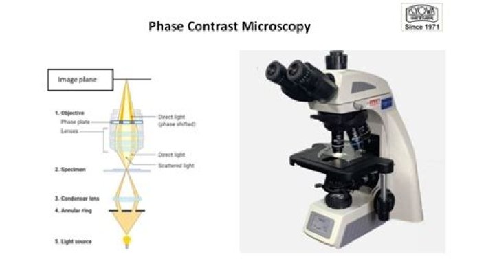
A phase plate is mounted in or near the objective rear focal plane (see Figures 4 and 5) in order to selectively alter the phase and amplitude of the surround (or undeviated) light passing through the specimen.
What is the principle of phase contrast microscopy?
Principle of Phase contrast Microscopy When light passes through cells, small phase shifts occur, which are invisible to the human eye. In a phase-contrast microscope, these phase shifts are converted into changes in amplitude, which can be observed as differences in image contrast.What condenser setting value do you want when you are using 100x objective lens?
Most condenser lens systems for research grade microscopes (1000x) have an N.A. value of 1.25. The 100x objective lens is also rated at 1.25. The medium between the 100x lens and the slide can be air or oil.
How does phase contrast microscopy differ from bright field microscopy?
Phase contrast is preferable to bright field microscopy when high magnifications (400x, 1000x) are needed and the specimen is colorless or the details so fine that color does not show up well. In phase contrast a phase plate is placed in the light path.
What type of microscopy is phase contrast microscopy?
Phase-contrast microscopy (PCM) is an optical microscopy technique that converts phase shifts in light passing through a transparent specimen to brightness changes in the image. Phase shifts themselves are invisible, but become visible when shown as brightness variations.
What does 10 0.25 mean on a microscope?
10/0.25: 10 = magnification (10x) 0.25 = numerical aperture.Which lens is used in phase contrast microscope?
When the condenser annulus and objective phase plate are in proper alignment, the image illustrated in Figure 1(e) should appear in the phase telescope or eyepiece with a Bertrand lens in place. At this point, the microscope is properly configured for observation of the specimen with phase contrast illumination.
How do you find the limit of resolution?The Rayleigh criterion stated in the equation θ=1.22λD θ = 1.22 λ D gives the smallest possible angle θ between point sources, or the best obtainable resolution. Once this angle is found, the distance between stars can be calculated, since we are given how far away they are.
Article first time published onWhat is the difference between 4x 10x and 40x on a microscope?
4x is a common magnification for scanning objectives and, when combined with the magnification power of a 10x eyepiece lens, a 4x scanning objective lens gives a total magnification of 40x.
How do you align a phase ring?
To ensure proper phase ring/plate alignment, first toggle the Phase Telescope radio button to the In position and use the sliders to align the plate outline within the phase ring. Then toggle the Phase Telescope out of the light path to view the specimen under optimum conditions of phase contrast illumination.
How do you take good pictures under a phase contrast microscope?
Thus, in order to achieve high-quality phase-contrast images, the correct phase plate and condenser annulus pair must be used and the condenser annulus must be properly centered such that the image of the annulus corresponds exactly with the position of the phase ring.
What methods are used to improve contrast in bright field microscopy?
Phase contrast microscopy is a technique used to increase contrast within transparent samples such as cells or thin tissue sections. The technique works by using an annulus at the condenser to only allow certain phases of light though the condenser and onto the sample.
What do phase contrast and dark field microscopy have in common?
Dark field and phase contrast microscopes allow to observe transparent samples. The dark field microscope produces a light cone, which reaches the objective only when it is scattered by the sample. … Making these two parts interfere creates a contrast, which allows to visualize samples even when they are transparent.
Is a phase contrast microscope an electron microscope?
Phase-contrast imaging is a method of imaging that has a range of different applications. … In transmission electron microscopy (TEM), phase contrast enables very high resolution (HR) imaging, making it possible to distinguish features a few Angstrom apart (at this point highest resolution is 40 pm).
What is the magnification of phase contrast microscopy?
Phase contrast is preferable to bright field microscopy when high magnifications (400x, 1000x) are needed and the specimen is colorless or the details so fine that color does not show up well. Cilia and flagella, for example, are nearly invisible in bright field but show up in sharp contrast in phase contrast.
What is a phase contrast condenser?
Phase Contrast Condenser: The condenser will contain what is called the annulus. The annulus is the second component to complete the optical aspect of phase contrast. The annulus must match with the phase plate (ring) inside of the phase contrast objective.
What does 40 0.65 mean on a microscope?
Roughly, a 40/0.65 objective lens delivers a blurrier image than a 40/1.3 objective lens. Note that the numerical aperture value is less significant at low magnifications. It should be taken into account only when choosing an objective lens with 40x magnification and higher.
What does 40x mean on a telescope?
Magnification = Telescope focal length ÷ Eyepiece focal length. For example, if you use a telescope of 1000mm focal length with a 25mm eyepiece, the magnification would be 40x (1000mm ÷ 25 = 40). Doubling the power gives you one-fourth the image brightness and reduces the sharpness by one half.
What does the 40 on this microscope objective lens mean?
Coverslips with a deviating thickness will result is an image of lower resolution. 4, 10, 20, 40, 100: This represents the magnification of the objective. The total magnification is calculated by multiplying the magnification of the objective with the magnification of the ocular (eye piece), which is usually 10x.
How do I increase resolution limit?
The resolution of a specimen viewed through a microscope can be increased by changing the objective lens. The objective lenses are the lenses that protrude downward over the specimen. Grasp the nose piece. The nose piece is the platform on the microscope to which the three or four objective lenses are attached.
How does wavelength affect resolution of a microscope?
Microscope resolution is also impacted by the wavelength of light being used to illuminate the specimen. Longer wavelengths of light offer less resolution than short wavelength illumination. … As light slows down the wavelength gets shorter and yields better resolution.
When switching to the 100x what should you use?
When switching to the 100x lens, what should you use? 100x lenses should be used with a few drops of immersion oil to enhance the image.
What magnification do I need to see bacteria?
While some eucaryotes, such as protozoa, algae and yeast, can be seen at magnifications of 200X-400X, most bacteria can only be seen with 1000X magnification. This requires a 100X oil immersion objective and 10X eyepieces.. Even with a microscope, bacteria cannot be seen easily unless they are stained.
How do you determine total magnification when using the 4X 10X and 40X objectives with a 10X ocular?
To figure the total magnification of an image that you are viewing through the microscope is really quite simple. To get the total magnification take the power of the objective (4X, 10X, 40x) and multiply by the power of the eyepiece, usually 10X.
How do you align a microscope?
To align, turn the brightness knob down to a fairly low setting, then remove the frosted glass filter from the light path. On an upright microscope place a piece of lens paper over the field diaphragm to see the image of the filament.
What is a condenser annulus?
The condenser annulus (illustrated in Figure 1) is typically constructed as an opaque flat-black (light absorbing) plate with a transparent annular ring, which is positioned in the front focal plane (aperture) of the condenser so the specimen can be illuminated by defocused, parallel light wavefronts emanating from the …
What kind of microscope is used for fluorescence imaging?
Most of the fluorescence microscopes used in biology today are epi-fluorescence microscopes, meaning that both the excitation and the observation of the fluorescence occur above the sample. Most use a Xenon or Mercury arc-discharge lamp for the more intense light source.
What are the two things that can be done to improve contrast?
Contrast may be improved by placing suitable apertures or filters within the optical path, either in the illuminating system alone (dark ground or Rheinberg illumination), or in conjugate planes in the imaging system (e.g. for phase contrast, differential interference contrast or polarised light microscopy).
How does a DIC microscope increase contrast?
Compensators in DIC Microscopy Specimen contrast can also be increased by introducing a retardation plate (or compensator) into the optical pathway in a DIC microscope.